If you’ve recently experienced a painful twist or strain in your knee—perhaps while playing sports, climbing stairs, or even walking—there’s a possibility you might have injured your meniscus. The meniscus is a vital piece of cartilage that cushions your knee joint, and a tear in this area can cause swelling, instability, or a locking sensation.
At the Dr. Rami Hamed Center (DRHC) in Dubai, many patients come to us worried about knee pain and unsure about the next steps. One of the most effective tools for diagnosing a meniscus tear is an MRI scan, which provides a clear, detailed image of the structures inside your knee, without the need for surgery.
In this post, we’ll explain how MRI helps diagnose meniscus tears, what to expect during the scan, and how we guide you through your recovery options.
What Is a Meniscus Tear?
Your knee contains two menisci—crescent-shaped cartilage discs that act as shock absorbers between the thigh bone (femur) and shin bone (tibia). A meniscus tear can happen suddenly due to trauma or twisting movements, or it can occur gradually with age-related degeneration.
Common signs of a meniscus tear include:
- A popping sensation at the time of injury
- Swelling or stiffness around the knee
- Difficulty bending or straightening the leg fully
- Knee locking or catching
- Pain during twisting or squatting motions
While a physical exam gives clues about the injury, an MRI is often needed to confirm the diagnosis, especially if surgery is being considered.
Why MRI Is the Gold Standard for Diagnosing a Meniscus Tear
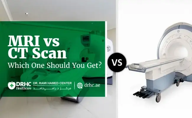
MRI (Magnetic Resonance Imaging) is a non-invasive imaging method that uses strong magnets and radio waves to create detailed cross-sectional images of the knee. It allows doctors to assess soft tissues like the meniscus, ligaments, cartilage, and tendons—structures that are not visible on X-rays.
An MRI can:
- Accurately identify the location and size of the tear
- Detect associated injuries such as ligament damage or fluid buildup
- Help determine whether conservative treatment or surgery is needed
At DRHC Dubai, our orthopedic and radiology teams work together to ensure timely diagnosis and a customized treatment plan based on your MRI results.
What to Expect During a Knee MRI in Dubai
We understand that getting an MRI can feel intimidating, especially if it’s your first time. Here’s what you can expect:
Before the Scan
- No special preparation is needed for a knee MRI.
- You can eat and take your medications as usual.
- Let the staff know if you have any metal implants, or pacemakers, or experience claustrophobia.
During the Scan
- You’ll lie comfortably on a flat table that slides into the MRI machine.
- The knee will be positioned carefully to ensure clear imaging.
- The machine is loud, but you’ll be given earplugs or headphones.
- The scan takes about 20–30 minutes.
- There’s no pain involved, but staying still is important for image clarity.
After the Scan
- You can resume normal activities immediately.
- The images are reviewed by a radiologist and shared with your orthopedic specialist.
- Based on the findings, we’ll discuss the next steps—from physical therapy to possible surgical repair.
Explore Our Related Blogs
- Will You Need Spine Surgery? Here’s How We Decide
- How Spine Surgery Has Changed in the Last 10 Years
- Understanding Endoscopic Spinal Decompression: Who It Helps
- What to Expect During Recovery from Endoscopic Spine Surgery
- Lower Back Pain That Won’t Go Away? You Might Need a Spine MRI
- MRI for Knee Pain – When It’s Time to Get Scanned
- Shoulder Pain That Won’t Go Away? You May Need an MRI
- MRI for Spinal Pain in Dubai – Lumbar vs Cervical
Frequently Asked Questions
Is the MRI painful?
No. MRI is completely non-invasive and pain-free. You may feel a bit stiff from holding still, but there’s no discomfort from the scan itself.
Can I walk on a torn meniscus?
In some cases, yes. But walking may worsen the tear or cause more damage, especially if the injury isn't diagnosed properly. Early MRI imaging helps prevent further joint problems.
Is surgery always required?
Not necessarily. Small tears or degenerative changes can often be managed with rest, physiotherapy, and anti-inflammatory medications. However, larger tears or those causing mechanical symptoms (like locking) may need arthroscopic surgery.
Is MRI covered by insurance in Dubai?
Many health insurance providers in the UAE cover MRI scans when they are medically indicated. Our team at DRHC will assist with approvals and guide you through the process.
When Should You See a Knee Specialist?
If your knee pain persists for more than a few days, or if you notice instability, swelling, or limited motion, it’s important to consult an orthopedic specialist. Delaying treatment could lead to worsening symptoms or even long-term joint damage.
At DRHC Dubai, we provide comprehensive orthopedic care, including advanced imaging like MRI, minimally invasive treatments, and rehabilitation programs tailored to your needs.
Final Thoughts: Taking the First Step Toward Healing
Living with knee pain can limit your ability to move, work, and enjoy everyday life. A meniscus tear, while common, deserves timely attention. An MRI scan is a safe, effective, and reliable way to understand the cause of your knee discomfort and find the best path forward.
Whether you're newly injured or still dealing with chronic knee pain, you don’t have to go through it alone. Consultations and imaging services are available at DRHC Dubai, where our specialists are ready to guide you through diagnosis, treatment, and full recovery.
📞 +971 4 279 8800
🌐 www.drhc.ae
📍 Dubai Healthcare City, Building 52
Topic: Radiology orthopedic MRI


Leave a comment