

.jpg?width=838&name=shutterstock_1023401932%20(1).jpg)
How a Laparoscopic Hernia Repair is done?
There are two Laparoscopic techniques for the surgical procedure of Inguinal Hernias.
- TEP (totally extra-peritoneal) repair: in which the approach is done through the abdominal wall only (pre-peritoneal) behind the rectus muscle.
- TAPP (trans-abdominal pre-peritoneal) repair: in which the approach is performed through the abdominal cavity.
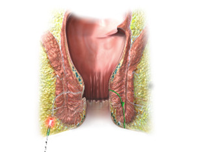
TEP (totally extra-peritoneal) repair:
Description of the procedure:
- The patient is placed in the supine position. Induction of General Anaesthesia.
- Insertion of a foley catheter is done aseptically. Disinfection and covering of abdomen according to standards.
- The first access is carried out through an infra-umbilical or para-umbilical incision of the skin until reaching the fascia.
- Preperitoneal space is entered after the elevation of the rectus muscle.
- Placement of Bilateral balloon trocar into the space behind the rectus muscles. Insufflation under direct visualization and the removal of the bilateral balloon are also under visualization.
- A 10 mm trocar is inserted into the pre-peritoneal space.
- Insufflation of this space with CO2 gas to reach the maximum pressure of 10-12 mmHg using a flow rate of 3-4 L/min.
- Placement of two 5 mm trocars 5cm away from the umbilicus towards the pubis symphysis respectively.
- Dissection of the peritoneum is carried out on both sides (pre-peritoneal).
- The hernia sac should be separated from the vas deferens and cord vessels in order to bring it into the pre-peritoneal sac. Then a 3D max MESH large size is brought and placed in a way to cover completely the internal ring.
- After deflation of the pre-peritoneal space, the peritoneum and fatty tissue will get together with the mesh avoiding displacement.
- All trocars are removed under direct visualization. Closure of the infra- or para-umbilical incision is carried out with interrupted or non-interrupted sutures. Closure of the skin incisions. Removal of foley catheter. Wound dressing.
End of operation.
TAPP (trans-abdominal pre-peritoneal) repair.
Description of the procedure:
After general anesthesia is induced.
- Insertion of foley catheter aseptically.
- The abdomen is prepped and draped sterilely. A small sub-umbilical incision is carried through skin and subcutaneous tissues until fascia is reached and dissected in a way to reach the peritoneum to get access to the abdominal cavity under visual control.
- This is the open technique, otherwise gaining access to the abdominal cavity can be also done with the v needle. Insufflation of the abdomen cavity to establish pneumo-peritoneum at a pressure of about 15 mmHg.
- The first port is placed sub-umbilical, the second port is para-rectal on the right side and the third port is placed para-rectal on the left side. Exploring the site of operation. In 10-15% of unilateral hernias cases, an intra-operative contralateral hernia can be found at the time of surgery.
- The peritoneum is incised from the anterior upper iliac spine to the medial umbilical ligament above the inner ring. The hernia sac is brought into the abdomen and completely reduced.
- The inguinal canal is explored and lipomas, if present is extracted and resected. After then, non-absorbable lightweight MESH is brought and adapted to underlying tissues and should be fixed by staples, absorbable or non-absorbable sutures, or tags with fibrin or glue.
- In order to prevent any bowel obstruction or direct contact with MESH, peritoneal closure is performed with running absorbable sutures.
- Removal of trocars under visual control while deflating the abdominal cavity. Bigger trocar wounds are closed in layers.
- Removal of foley catheter. Wound dressing.
End of operation.
Laparoscopic Hernia Repair is also available for Incisional, Umbilical, or Hiatal Hernia with low recurrence and complication rates.
Description of the procedure:
- The patient is placed in the supine position. After general anesthesia is induced. The abdominal skin is sterilized and draped.
- A pneumo-peritoneum is achieved with the needle or in an open technique. Ports are placed according to the site of the Hernia.
- A laparoscopic examination of the abdomen is performed and any abnormalities would be noted. If there is no contraindication to proceed, the incarcerated contents are reduced.
- The undersurface of the abdominal wall is cleared from any fatty deposits which may inhibit the flat application of the MESH.
- The fascial defect is closed with absorbable sutures. The MESH is then rolled and inserted through the port into the abdominal cavity.
- The MESH is unrolled inside the abdomen and positioned against the abdominal wall. It is important to have a 3-5 cm overlap over the entire fascial defect.
- Fixation of the MESH on the undersurface of the abdominal wall is achieved either with tuckers or extra sutures.
- Control of placement is performed. Deflation of the abdominal cavity while removing the trocars.
- Closure of trocar wounds.
End of operation.
This may be so many technical points, but many of my patients have asked very detailed questions about the procedure, hence I thought to put it in writing if they wish to read it in full detail.
Read
- Experience and vision of Laparoscopic Hernia Repair
- Open VS Laparoscopic Hernia repair.
- 20 Q&A about Laparoscopic Hernia Repair
- Latest techniques for Laparoscopic Hernia Repair
- Possible complications after Laparoscopic Hernia Surgery
- Recovery after Laparoscopic Hernia Repair
- Best practices in Hernia Surgery Repair
*Find out more about Laparoscopic Hernia surgery Here*

*Laparoscopic Hernia surgery Cost*

.jpg)

.png)

Leave a comment